Figure template · Mechanism & Pathway · 4:3
PD-1/PD-L1 Checkpoint Contacts in the Tumour Microenvironment Figure
A T cell at the centre meets a tumour cell at two molecular contacts, PD-1 with PD-L1 above and TCR with MHC below, while an anti-PD-1 antibody and a blood vessel set the scene for checkpoint immunotherapy papers and talks.
Prompt
Create a scientific figure about: PD-1 / PD-L1 checkpoint contacts in the tumor microenvironment. Visual style: Clean Vector. Requirements: publication-ready scientific quality, clear labels and directional arrows where appropriate, avoid cartoon or stock-photo look, composition optimized for research papers and academic slides.
What this template is
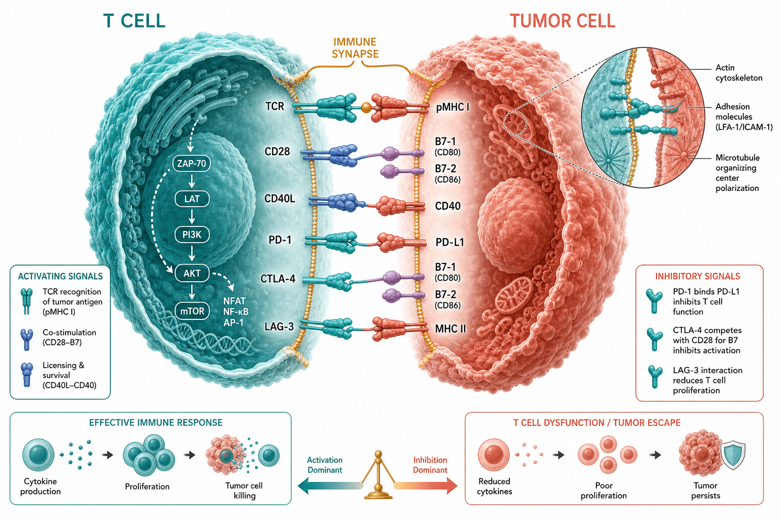
The figure zooms in on one encounter inside a tumour. The T cell recognises antigen presented by MHC on the tumour cell through its TCR, while PD-1 on the same T cell engages PD-L1 on the tumour cell and delivers an inhibitory signal. An anti-PD-1 antibody hovering above the contact stands for checkpoint blockade. A vessel lined with endothelial cells, neighbouring T cells and tumour cells, and scattered small particles give the microenvironment around them. A reader should come away understanding why blocking this receptor–ligand pair can let the T cell resume killing.
Elements to label
T cell
The central teal cell with a leader line to its body; specify CD8⁺ cytotoxic T cell if that is your subject.
Tumour cell
The large pink cell at right, studded with surface molecules; its label sits in clear space inside the cell.
PD-1 and PD-L1
The upper contact, where a blue molecule from the T cell meets PD-L1 on the tumour cell; label each partner separately.
TCR and MHC
The lower contact with a purple antigen peptide between TCR and MHC; add the MHC class to the label.
Anti-PD-1 antibody
A teal Y-shaped molecule above the cells with a dashed arrow and an inhibition bar; a few more float in the background.
Blood vessel and endothelium
A curved vessel at left containing red cells, with endothelial cells labelled, hinting at immune cell entry into the tumour.
Cytokine particles
Unlabelled teal and purple dots drifting around the T cell; name them as interferon or whichever factors your data support, or remove them.
Where it is used
- Paper mechanism figure
- Graphical abstract
- Defense slides
- Grant proposal
What reviewers look for
PD-1 drawn like an antibody
PD-1 is a receptor with a single immunoglobulin-like domain, but the sample draws it as a Y-shaped molecule almost identical to the therapeutic antibody. Ask for a single-chain receptor so the two cannot be confused.
Blockade logic
An anti-PD-1 antibody should sit on PD-1 at the T cell surface, preventing PD-L1 binding. In the sample it floats free and its inhibition bar points toward the tumour side, which reads more like an anti-PD-L1 drug.
No downstream consequence
Reviewers expect to see PD-1's cytoplasmic tail recruiting SHP-2 to damp TCR and CD28 signalling, and restored T cell function after blockade. The sample shows no signalling outcome at all.
Cell identity and antigen presentation
State the T cell subset and the MHC class, since CD8⁺ T cells recognise MHC class I. The peptide and the background particles are unlabelled in the sample, so readers cannot tell what they represent.
How to use it in Yila
- 1Pick a figure
- 2Describe your subject and the steps to stress
- 3Refine it in chat, then download the PNG
Tips
Do
- State whether your drug targets PD-1 or PD-L1 so the antibody is drawn bound to the right molecule.
- Ask for PD-1 as a single-chain receptor in a colour distinct from the antibody, so two Y shapes never sit side by side.
- If your work concerns exhaustion, ask for an arrow from exhausted to reinvigorated states beside the T cell, labelled with the markers you measured.
Avoid
- Leaving floating antibodies and cytokine dots unlabelled; readers will guess at what they mean, and reviewers will ask.
- Using the glossy rendered style at column width, where the textured cell surfaces turn into visual noise and labels lose contrast.
- Dropping the TCR–MHC contact; a checkpoint-only drawing hides the activation step that checkpoints regulate.
Questions
Can this checkpoint figure go straight into my paper?
Use it as a draft. It is AI-generated, and both the shape of PD-1 and the position of the antibody need correcting. Check every molecule name and the direction of each interaction against your study before it becomes a formal figure; the biology remains your responsibility.
Can elements in the figure be changed, such as molecules or cells?
The figure is not editable element by element and is not a vector graphic. Tell Yila in chat to switch to an anti-PD-L1 antibody, add myeloid suppressor cells or enlarge the contact zone, and it regenerates the whole image with those changes in place.
What format is delivered?
An AI-generated PNG in landscape orientation that drops straight into slides or documents. When combining it with flow cytometry or imaging panels, set matching font sizes in your layout software before exporting the final multi-panel figure.
Is it free?
Browsing this page and the sample costs nothing. Generating a figure in Yila, and revising it, consumes credits. New accounts receive 2,500 credits, enough to produce a first version and judge whether it suits your immunotherapy project before you invest in further rounds.
Can it add regulatory T cells or macrophages to the microenvironment?
Yes. Name the cell types and how they relate to the tumour or the T cell, for example by secreting suppressive factors, and Yila places them in the background with labels so the composition of the microenvironment becomes explicit rather than generic.
Can it show a combination therapy?
Describe the partner drug and its target, such as a second checkpoint or a targeted agent. Yila can add the second antibody or drug on the relevant cell and use arrows to show how the two treatments are thought to cooperate, which is useful for grant proposals.