Figure template · Mechanism & Pathway · 4:3
NF-κB Inflammatory Activation in Macrophages
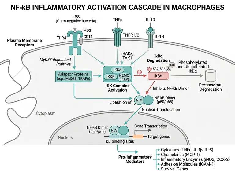
A landscape macrophage figure in which three inflammatory stimuli converge on the IKK complex, IκBα degradation frees the p50/p65 dimer to enter the nucleus, and the resulting cytokines, chemokines and enzymes are listed below, for inflammation and immunology papers.
Prompt
Create a scientific figure about: NF-κB inflammatory activation cascade in macrophages. Visual style: Clean Vector. Requirements: publication-ready scientific quality, clear labels and directional arrows where appropriate, avoid cartoon or stock-photo look, composition optimized for research papers and academic slides.
What this template is
LPS is handed by CD14 and MD2 to TLR4, while TNFα and IL-1β bind their own receptors. Signals pass through MyD88, TRAF6, the IRAKs and TAK1 to the IKK complex of IKKα, IKKβ and NEMO. IKK phosphorylates IκBα at S32 and S36, the inhibitor is ubiquitinated and destroyed by the proteasome, and the p50/p65 dimer exposes its nuclear localization signal, moves into the nucleus and binds κB sites. Transcription of TNFα, IL-6, MCP-1, iNOS, COX-2 and ICAM-1 follows. A reader should grasp that destroying the inhibitor is the switch.
Elements to label
Three stimuli and receptors
LPS with TLR4, CD14 and MD2; TNFα with its receptor; IL-1β with IL-1R, spread across three columns at the membrane.
MyD88-dependent route
An adaptor box naming MyD88 and TRAF6, with arrows labelled as coming from TLR4 and IL-1R.
IRAKs and TAK1
Place them where the three receptor signals merge, captioned as the kinases acting upstream of IKK activation.
IKK complex
IKKα, IKKβ and NEMO in one box, with NEMO also labelled IKKγ.
IκBα phosphorylation
Mark the S32 and S36 sites and the ubiquitin chain; the sample writes a Latin k where the Greek letter belongs.
Proteasomal degradation
An arrow from IκBα into the proteasome, captioned to say degradation is what releases NF-κB.
p50/p65 dimer
The dimer in the cytoplasm with its NLS exposed, then again in the nucleus aligned over κB binding sites.
Pro-inflammatory outputs
Cytokines, chemokines, inflammatory enzymes, adhesion molecules and survival genes listed on separate lines.
Where it is used
- Paper mechanism figure
- Graphical abstract
- Defense slides
- Course handout
What reviewers look for
How IκBα meets the dimer
The sample draws an ordinary arrow from IκBα to the dimer while the caption says it inhibits. At rest IκBα holds p50/p65 in the cytoplasm, so show binding or a blunt line and make clear that release follows degradation.
Adaptors cannot bypass the IKK complex
One arrow runs from the adaptor box straight to NF-κB. In canonical signaling MyD88 and TRAF6 act through TAK1 and IKK before the dimer is freed, so a reviewer will ask for that shortcut to go.
Use the Greek kappa, not a Latin k
The title and several labels spell NF-kB and IkBα with a Latin k. Immunology editors and reviewers expect the Greek κ throughout, in the figure as well as the text.
TLR4 has a second route
After endocytosis TLR4 signals through TRIF to late NF-κB activation and interferon responses. A paper comparing MyD88 and TRIF should add the endosomal TRIF branch the sample leaves out.
How to use it in Yila
- 1Pick a figure
- 2Describe your subject and the steps to stress
- 3Refine it in chat, then download the PNG
Tips
Do
- State your stimulus and time point, for example LPS alone, so Yila removes the two receptor inputs you did not use.
- If you imaged p65 nuclear translocation, ask for a phosphorylated p65 mark in the nucleus that matches your immunofluorescence data.
- Ask for a dashed IκBα resynthesis arrow from the nucleus if your time course shows the negative feedback that ends the response.
Avoid
- Do not keep the Latin NF-kB spelling from the sample; check the title and every label after each round.
- Do not list more mediators than you measured; if you assayed only IL-6 and iNOS, keep the output box to those.
Questions
Can this NF-κB figure go into my paper unchanged?
Use it as a first draft of the mechanism panel. It is an AI-generated image that spells kappa with a Latin k, draws an unclear arrow between IκBα and the dimer and adds an adaptor shortcut around IKK, so fix those points and keep refining it in chat before submission.
Can I switch the macrophage for microglia or epithelial cells?
Not element by element, but you can keep refining it by chatting in Yila. Name the cell type and the receptors it expresses, for example microglia with TLR4 only, and it will rewrite the title, receptors and output mediators before generating a new version for you to check.
What format is the output?
An AI-generated PNG image in a 4:3 landscape frame, ready for a figure panel or defense slide. It is not a vector graphic, and the phosphorylation sites and ubiquitin marks are small, so check them at your final print size before enlarging the figure, and ask for bigger labels if they fade.
Does generating it cost anything?
Opening this page and looking at the sample cost nothing. Generating and editing in Yila consumes credits, and a new account receives 2,500 credits, enough for several versions tuned to your own stimulation conditions, cell type and readouts.
Can I add the inflammasome and IL-1β maturation?
Yes. Ask for NF-κB to be drawn as the priming signal and for a second step showing NLRP3 inflammasome assembly, caspase-1 activation and cleavage of the IL-1β precursor, so both signals of the two-step model share one figure and the link between transcription and secretion is visible.
Can it mark where my inhibitor acts?
Tell it which step your compound targets, such as IKKβ or the proteasome, and it will place an inhibition symbol and the drug class at that node. That makes the figure easy to read alongside your pharmacology experiments and shows reviewers exactly which part of the cascade you blocked.