Figure template · Mechanism & Pathway · 4:3
T-cell Immune Synapse and Checkpoint Figure
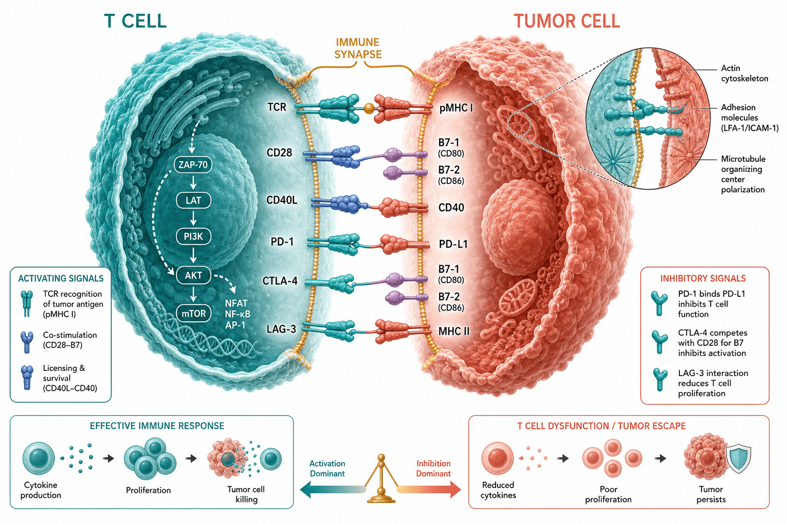
Two cells face each other across the synaptic cleft with receptors paired row by row, activating signals running into the T cell and inhibitory contacts listed apart, and a balance at the foot of the figure weighing tumour killing against immune escape.
Prompt
Create a scientific figure about: T-cell and tumor immune synapse with checkpoint contacts. Visual style: Clean Vector. Requirements: publication-ready scientific quality, clear labels and directional arrows where appropriate, avoid cartoon or stock-photo look, composition optimized for research papers and academic slides.
What this template is
The middle of the figure is the synapse itself: the two membranes come close and receptors meet their ligands in one horizontal row each, so antigen recognition, co-stimulation and checkpoint contacts can be read in order. Inside the T cell a kinase cascade runs from recognition down to the transcription factors, while an inhibitory panel sits on the tumour side. Two small sequences along the bottom turn those molecular events into outcomes, cytokine release, proliferation and killing on one side against poor proliferation and a surviving tumour on the other, with a balance in the middle showing which of the two sides dominates.
Elements to label
T cell side
A cut-open cell on the left with its receptors facing the cleft and open space inside for the signalling cascade.
Tumour cell side
The right-hand cell carries only its ligands and a nucleus, in a colour clearly different from the T cell.
Synaptic cleft
Keep a narrow labelled gap between the two membranes and place every receptor pair on that same line.
Antigen recognition pair
The top row shows the receptor reading the peptide-loaded molecule, marked as the start of the whole cascade.
Checkpoint contacts
Give PD-1 with PD-L1, CTLA-4 and LAG-3 one row each, drawn in a style unlike the activating pairs.
Intracellular cascade
Arrows run from top to bottom through the kinases and end at the transcription factors, never curving into a loop.
Adhesion inset
A circular zoom at the upper right naming LFA-1 and ICAM-1, the actin cortex and the polarized organizing centre.
Outcomes and balance
Two short sequences end in tumour killing and in tumour persistence, with the balance tipping between them.
Where it is used
- Paper mechanism figure
- Defense slides
- Grant proposal
- Course handout
What reviewers look for
Ligands on the right cell
Checkpoint ligands belong to the tumour or myeloid cell and the receptors to the T cell; a swapped pair is the first thing a reader catches, and the adhesion molecules drawn inside the inset need exactly the same care.
Inhibition is not an off switch
Checkpoint contacts lower signal strength rather than cutting it, so draw a damped line rather than a broken one, and say in the caption whether it acts on recognition or on co-stimulation.
Cascade order is a claim
Readers treat the order of the intracellular arrows as a mechanistic statement, so leave out branches your text does not argue, and never string parallel transcription factors into one straight line.
Outcomes follow the data
Cytokine release, proliferation and killing should each map onto something you actually measured, and the way the balance tips needs experimental support rather than resting on the pathway sketch alone.
How to use it in Yila
- 1Pick a figure
- 2Describe your subject and the steps to stress
- 3Refine it in chat, then download the PNG
Tips
Do
- Keep only the receptor pairs your own study touches; the fewer rows crossing the cleft, the clearer the main line reads.
- Fix one colour per meaning in the prompt, such as recognition, co-stimulation and inhibition, and ask for it to hold across the whole figure.
Avoid
- Do not let the inset sit over the cleft; it should lead out into empty space so both layers stay readable.
- Do not draw the two outcomes as equal halves unless your own data really show both fates occurring about as often.
Questions
Can this go straight into a paper?
Use it as a draft of your mechanism panel. It is an AI-generated raster image, so check every row for the right receptor on the right cell, confirm the arrow directions and abbreviation styles against your text, and keep refining it in chat until the line weights and type sizes match what the journal asks for.
Can I change elements in the figure?
Not element by element, but you can keep refining it by chatting in Yila: remove the pairs you do not study, turn the tumour cell into an antigen-presenting cell, move the inset to the other corner, or keep only the activating side, then generate a new version from that description.
What format is delivered?
An AI-generated PNG image in landscape, suited to a mechanism panel, a defense slide or a teaching handout. It is not a vector graphic, and the small receptor labels soften when the figure is scaled down, so check it at the size you will actually print or project.
Is it free?
Browsing the page and viewing the sample cost nothing. Generating your own version in Yila consumes credits, and a new account receives 2,500 credits, which is enough to rework the receptor rows, the intracellular cascade and the outcome strip several times over.
I work on bispecific antibodies. Can it be adapted?
Yes. Keep the cleft and the two outcomes, replace one receptor row with your own molecule, and state in the side panel whether it bridges the two cells or blocks an existing contact, trimming away the rows your study does not need.
Can I keep only the synapse part?
Ask for the two cells and their paired contacts alone, without the outcome strip and without the inset. The result is sparser and works better as a single explanatory slide during a talk than the full figure does.