Figure template · Mechanism & Pathway · 4:3
Immune Synapse Figure with Receptor Contacts at the Interface
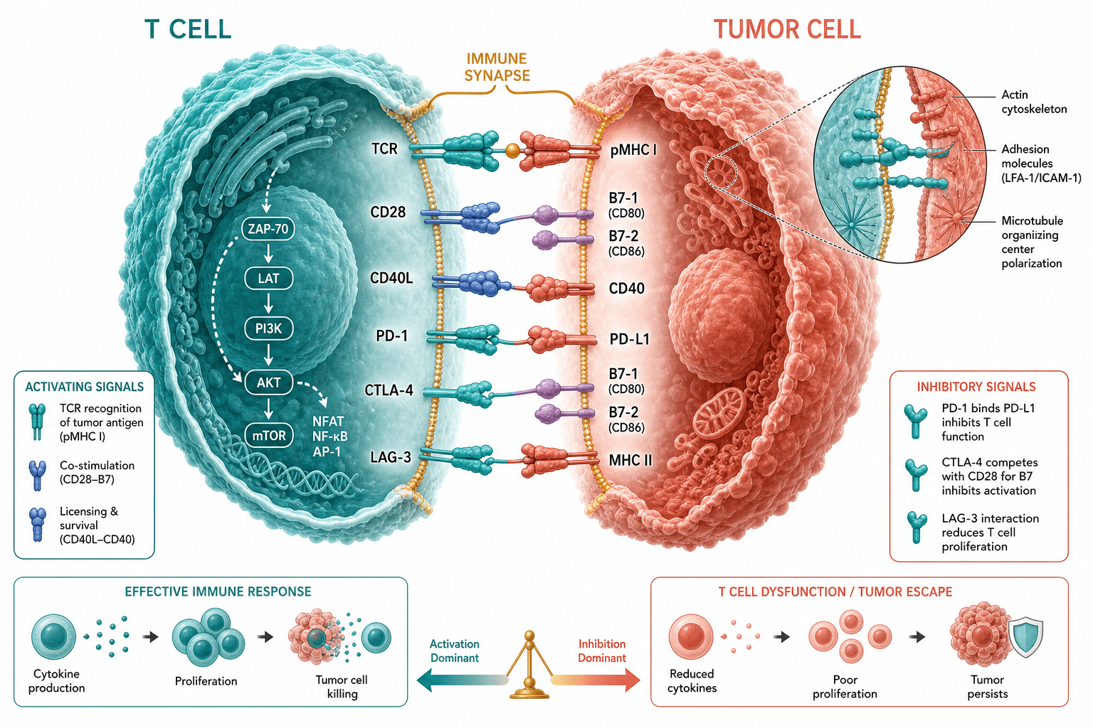
Two textured cells face each other while the gap between them is enlarged into rows of receptor-ligand contacts, with lytic granules and polarizing fibers inside the T cell, suited to a mechanism figure or an immunology lecture handout.
Prompt
Create a scientific figure about: Publication-style immune synapse mechanism figure highlighting receptor contacts at the cell interface. Visual style: Clean Vector. Requirements: publication-ready scientific quality, clear labels and directional arrows where appropriate, avoid cartoon or stock-photo look, composition optimized for research papers and academic slides.
What this template is
The figure zooms in on the contact zone between a T cell on the left and a target cell on the right. Across the interface, molecular pairs are laid out row by row: antigen recognition by the T-cell receptor and peptide-MHC, then costimulation through CD28 and B7, adhesion through LFA-1 and ICAM-1, and CD40L with CD40. Inside the T cell, lytic granules and fibers radiating toward the interface suggest the secretory machinery polarizing to the synapse, and clusters of small spheres mark cytokine release. A reader should be able to name the molecular pairs that form and stabilize the synapse and see the granules turned toward the target.
Elements to label
T cell
The large blue cell at the left, labelled from outside the upper corner with a leader line that touches the surface, not the nucleus.
Target cell
The pink cell at the right; in a final figure, say whether it is a tumor cell, an infected cell or an antigen-presenting cell.
TCR and pMHC
The top row, antigen recognition, with T-cell blue and target-cell orange showing which side each molecule belongs to.
Costimulation and adhesion pairs
CD28 with B7 and LFA-1 with ICAM-1 on separate rows, each name aligned on its own side of the gap.
CD40L and CD40
The bottom pair; for a study of cytotoxic killing, consider swapping it for a molecule relevant to that process.
Lytic granules
Textured spheres in the upper and lower T cell; one label with a single leader line is enough.
Microtubule organizing center
The point where the radiating fibers converge; its leader line must end there, not on the nucleus.
Cytokines
Distinguish the pro-inflammatory cluster above from the secreted spheres below by color, and state which cell releases each.
Where it is used
- Paper mechanism figure
- Course handout
- Academic poster
- Defense slides
What reviewers look for
The organizing center is mislabelled
In the sample, the leader line for the microtubule organizing center ends on the large sphere at the middle of the T cell, which reads as the nucleus; the center should be a small point between nucleus and interface, where the fibers start.
One T-cell type throughout
Lytic granules imply a cytotoxic T cell, while CD40L with CD40 is mostly a helper T cell talking to an antigen-presenting cell; showing both in one frame invites the question of which cell is drawn.
MHC class and co-receptor
The figure says pMHC without naming the class and draws no CD8 or CD4 co-receptor; reviewers expect a matched pair beside the recognition row, consistent with the T-cell type you describe.
Spatial arrangement
In a real synapse the receptor clusters at the center and LFA-1 forms an outer ring; four evenly spaced rows are a schematic shorthand, so add a face-on ring view if your paper discusses synapse structure.
How to use it in Yila
- 1Pick a figure
- 2Describe your subject and the steps to stress
- 3Refine it in chat, then download the PNG
Tips
Do
- Name the T-cell subset and the target-cell type in the prompt, so the pairs at the interface fit the cells drawn.
- For a clean vector look, ask in chat for flat fills and thin outlines in place of the textured cell surfaces.
- Ask for the receptor rows to follow the order your text discusses them, so the figure and the results section read in step.
Avoid
- Do not let leader lines cross other structures before landing; small targets such as the organizing center are the easiest to miss.
- Avoid drawing both cytokine clusters as identical colored spheres, or readers cannot tell which cell released which.
Questions
Can this synapse figure go straight into a paper?
Use it as a draft. It is an AI-generated raster image, and the sample already has at least one label pointing at the wrong structure; the molecular pairs and the cell types also need to match your study before it becomes a final figure.
Can the molecules and labels be changed?
Not element by element, but you can keep refining it by chatting in Yila: replace the CD40 row with PD-1 and PD-L1, add a CD8 co-receptor, move the organizing-center label to the right place, or remove the cytokine spheres, and then generate a new version.
What format is delivered?
An AI-generated PNG image in landscape, with textured, three-dimensional-looking cells. It is not a vector graphic, and the molecule names on either side of the gap are small enough to become hard to read at single-column width, so check them at the size you will use.
Is it free?
Viewing this page and the sample costs nothing. Generating your own version in Yila consumes credits; new accounts start with 2,500 credits, enough to try a few combinations of molecular pairs and then refine the one you prefer.
How does this differ from the simpler immune synapse figure?
This one shows only the two cells and the receptor pairs between them, with surface texture and granule detail, keeping the focus on the contacts themselves. It has no intracellular signalling cascade and no comparison of outcomes, so it fits when the interface molecules are the point.
Can it show a natural killer cell synapse instead?
Yes. Ask for the T cell to become a natural killer cell, change the recognition row to an activating receptor and its ligand, and add an inhibitory receptor pair; the lytic granules and the polarized structures facing the interface can stay.