Figure template · Mechanism & Pathway · 4:3
TGF-β/SMAD Signaling Pathway Figure
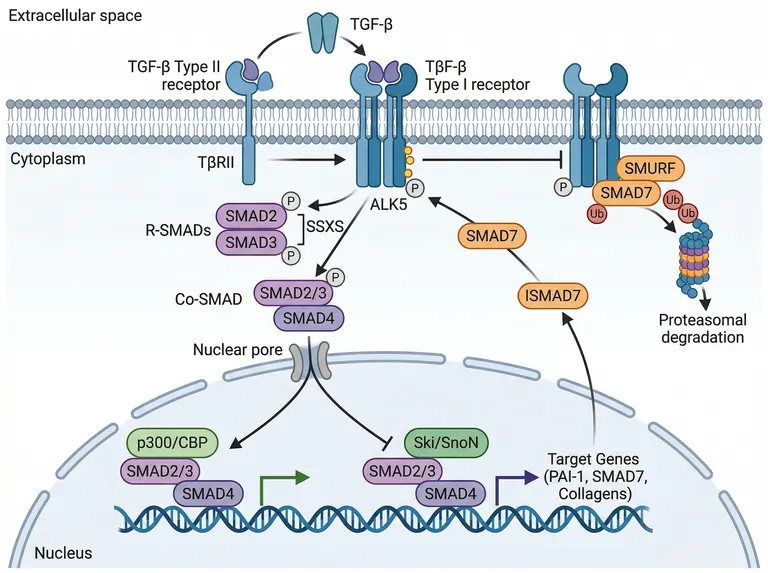
A landscape pathway figure layered as extracellular space, cytoplasm and nucleus, running from receptor activation to SMAD-driven transcription with SMAD7 feedback and receptor degradation on the right, for fibrosis, metastasis and EMT papers.
Prompt
Create a scientific figure about: TGF-β/SMAD pathway with receptor activation, R-SMAD phosphorylation, nuclear complex formation, and transcription. Visual style: Clean Vector. Requirements: publication-ready scientific quality, clear labels and directional arrows where appropriate, avoid cartoon or stock-photo look, composition optimized for research papers and academic slides.
What this template is
A TGF-β dimer binds the type II receptor, which recruits and phosphorylates the type I receptor ALK5. ALK5 phosphorylates the SSXS motif of SMAD2 and SMAD3; they partner with SMAD4, pass a nuclear pore and either join p300/CBP to switch on targets such as PAI-1 and collagens or meet the Ski/SnoN repressors. One target, SMAD7, returns to the membrane and, with SMURF, sends the receptor to the proteasome. The reader should see both the forward signal and the loop that shuts it off.
Elements to label
TGF-β ligand dimer
Place it at the top in extracellular space, with arrows to the type II receptor and then the receptor complex.
Type II and type I receptors
TβRII and ALK5 side by side across the membrane; the sample's type I label garbles the ligand name.
R-SMAD phosphorylation
SMAD2 and SMAD3 each carrying a phosphate, with a bracket naming the C-terminal SSXS motif.
SMAD complex
Stack the R-SMADs with SMAD4 and apply the Co-SMAD label to SMAD4 alone.
Nuclear pore and two outcomes
After the pore the path forks: one branch meets p300/CBP for activation, the other Ski/SnoN for repression.
Target genes
List PAI-1, SMAD7 and collagens beside the transcription arrow, noting that SMAD7 is itself a target.
SMAD7 feedback
Draw the returning SMAD7 with a blunt inhibitory end; the sample uses a pointed arrow and adds a stray ISMAD7 label.
SMURF and degradation
SMURF and SMAD7 bound to the receptor, with a ubiquitin chain leading to the proteasome.
Where it is used
- Paper mechanism figure
- Grant proposal
- Defense slides
- Course handout
What reviewers look for
Co-SMAD is on the wrong object
Co-SMAD means SMAD4 only, but the sample places the label beside the whole SMAD2/3 plus SMAD4 stack. Reviewers will ask for the R-SMAD and Co-SMAD to be labelled separately.
Feedback needs an inhibition symbol
SMAD7 competes for ALK5 and blocks R-SMAD phosphorylation, yet the sample draws an activating arrow from SMAD7 to ALK5. That says the opposite of the biology and must become a blunt line.
Complex stoichiometry
The active complex is usually described as a trimer of two R-SMADs with one SMAD4. If your text discusses assembly or nuclear retention, state the composition in the legend instead of showing a simple pair of stacked shapes.
Non-canonical branches
TGF-β also acts through TAK1, p38 and PI3K outside the SMAD route. If your EMT study measured these, add a side branch, or state in the legend that only canonical signaling is drawn.
How to use it in Yila
- 1Pick a figure
- 2Describe your subject and the steps to stress
- 3Refine it in chat, then download the PNG
Tips
Do
- Name the targets you measured, such as SNAIL or fibronectin, so the nuclear transcription box matches your results.
- For BMP work, describe the different ligands, receptors and R-SMADs so Yila renames the whole cascade consistently.
- Ask for the receptor inhibitor you used to be drawn on ALK5 with a blunt line if your paper tests pharmacological blockade.
Avoid
- Do not keep the garbled receptor labels; check the TβRII and ALK5 spellings in every regenerated version before export.
- Do not draw forward signaling and feedback with the same arrowhead; activation and inhibition need different line ends.
Questions
Can I submit this TGF-β figure as it is?
Use it as a draft of your mechanism panel. It is an AI-generated image with a garbled type I receptor label, a Co-SMAD label on the wrong object and an activating arrow where SMAD7 should inhibit. Correct those three points first, compare the remaining labels with the papers you cite, and keep refining it in chat before it goes to a journal.
Can I add my inhibitor or switch to the BMP pathway?
Not element by element, but you can keep refining it by chatting in Yila: put a small-molecule inhibitor on ALK5, or rename the ligand, receptors and R-SMADs for a BMP system, then generate a new version and compare it with the first.
Which file format is delivered?
An AI-generated PNG image in a 4:3 landscape frame, suited to a figure panel or slide. It is not a vector graphic; the small phosphate circles and ubiquitin marks blur when the image is shrunk into a multi-panel figure, so check them at final size and ask for larger symbols if they disappear.
Is generating this figure free?
Viewing this page and the sample is free of charge. Generating and editing in Yila consumes credits, and a new account receives 2,500 credits, enough to swap in your own targets and regulators over several rounds of revision before you settle on the panel.
Can it show the dual role of TGF-β in cancer?
Ask for two columns in the conversation: growth suppression in early tumours on the left, EMT and invasion in late tumours on the right, both sharing the same receptor and SMAD elements so the contrast is about outcome rather than machinery. Then regenerate and adjust the balance between the two halves.
I study fibrosis. Can collagen deposition stand out?
Yes. Say you want collagen deposition and myofibroblast activation emphasised, and it will enlarge collagen and PAI-1 in the nuclear transcription box and can add a short sketch of matrix building up outside the cell. Mention the tissue you study, such as lung or liver, so the extracellular sketch fits your model.