Figure template · Mechanism & Pathway · 4:3
PI3K-AKT-mTOR Signaling Axis in Cancer Cell Survival
A landscape signaling figure that radiates from AKT at its centre, fed from above by growth factor receptors and PI3K and branching out to anabolism, apoptosis blockade, cell cycle progression and angiogenesis, for oncology papers on drug targets and resistance.
Prompt
Create a scientific figure about: PI3K-AKT-mTOR signaling axis in cancer cell survival. Visual style: Clean Vector. Requirements: publication-ready scientific quality, clear labels and directional arrows where appropriate, avoid cartoon or stock-photo look, composition optimized for research papers and academic slides.
What this template is
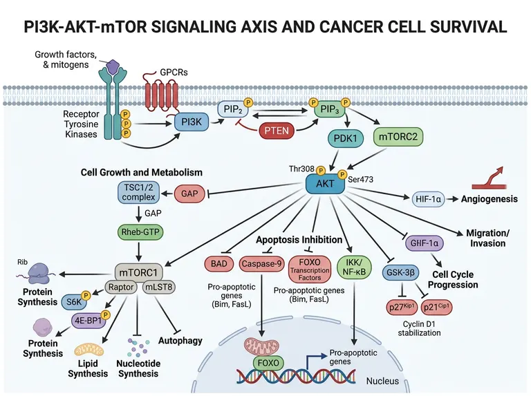
Growth factors activate receptor tyrosine kinases and GPCRs, and PI3K converts PIP2 to PIP3 while PTEN reverses that step. PIP3 brings PDK1 and mTORC2 to AKT, which they phosphorylate at Thr308 and Ser473. Active AKT relieves TSC1/2 restraint on Rheb so mTORC1 can drive protein, lipid and nucleotide synthesis through S6K and 4E-BP1 while suppressing autophagy, and it silences BAD, caspase-9 and FOXO to block apoptosis. Further branches run to GSK-3β, HIF-1α and migration. A reader should leave knowing why tumour cells lean on this axis to stay alive.
Elements to label
Receptors and upstream input
Receptor tyrosine kinases and GPCRs in the membrane under their growth factor ligands, with arrows converging on PI3K.
PIP2 and PIP3
Two lipids on the inner leaflet told apart by their phosphate count, with the PI3K arrow running from PIP2 to PIP3.
PTEN
It should act on PIP3 and return it to PIP2; the sample draws its arrow and its blunt end the wrong way round.
AKT dual phosphorylation
AKT at the centre, with Thr308 credited to PDK1 and Ser473 to mTORC2.
TSC1/2 and Rheb
AKT inhibits TSC1/2 and TSC inhibits Rheb; the sample shows the second step as activation.
mTORC1 outputs
Mark S6K and 4E-BP1 phosphorylation, lipid and nucleotide synthesis, and a blunt line onto autophagy.
Apoptosis blockade
BAD, caspase-9 and FOXO each joined to AKT by a blunt line, so the reader sees all three switched off.
Further outputs
GSK-3β, HIF-1α and migration each take a branch; one garbled label beside GSK-3β should be removed.
Where it is used
- Paper mechanism figure
- Grant proposal
- Defense slides
- Academic poster
What reviewers look for
PTEN points the wrong way
PTEN removes the 3-phosphate from PIP3 and returns it to PIP2, but the sample aims its blunt end at PIP2 and its arrowhead at PIP3. Any reviewer in the field will flag this reversal first.
The TSC and Rheb logic
The TSC complex is the GTPase-activating protein that switches Rheb off. Because the sample draws an activating arrow from TSC to Rheb, the idea that AKT relieves an inhibition no longer reads.
Where things sit in the cell
The sample places a mitochondrion inside the nucleus and runs the caspase-9 arrow into it. Apoptotic machinery belongs in the cytoplasm and mitochondria; only FOXO enters the nucleus, and only when AKT is not phosphorylating it.
The missing feedback loop
S6K downstream of mTORC1 feeds back to inhibit IRS1, the classic explanation for AKT reactivation under mTORC1 inhibitors. A paper about resistance should draw that loop explicitly.
How to use it in Yila
- 1Pick a figure
- 2Describe your subject and the steps to stress
- 3Refine it in chat, then download the PNG
Tips
Do
- Name your tumour type and lesion, such as an activating PIK3CA mutation or PTEN loss, so the figure marks the affected node.
- List the drugs you test against PI3K, AKT or mTORC1 so Yila adds inhibition symbols at exactly those nodes.
- Ask for the S6K to IRS1 feedback loop when your discussion turns to adaptive resistance under mTORC1 inhibition.
Avoid
- Do not keep the reversed PTEN and TSC arrows from the sample; check those two connections first in every version.
- Do not give every downstream branch equal weight when your paper discusses only one or two; fade the rest so readers find your result.
Questions
Can this PI3K-AKT-mTOR figure go straight into a paper?
Only as a starting draft. It is an AI-generated image in which the PTEN and TSC arrows run the wrong way, a mitochondrion sits inside the nucleus and one label is garbled. Correct those points against the literature you cite, then keep refining it in chat until it is ready to submit.
Can I keep only the branch I study?
Not element by element, but you can keep refining it by chatting in Yila. Ask it to keep only the mTORC1 anabolic branch and fade or drop the others, or to add the phosphorylation sites you measured by western blot, and then generate a new version and compare the two side by side.
What format do I download?
An AI-generated PNG image in a 4:3 landscape frame, suited to papers and defense slides. It is not a vector graphic, and small labels such as the phosphorylation sites soften when enlarged, so check legibility before using it at poster size or ask for larger type on the nodes you care about.
Do I pay to generate it?
Viewing this page and the sample costs nothing. Generating or revising in Yila consumes credits, and a new account receives 2,500 credits, enough to tailor several versions to your own mutations and drug targets before you choose one for the manuscript.
Can it show pathway blockade under combination therapy?
Yes. Describe the combination in the conversation, for example a PI3K inhibitor with a MEK inhibitor, and it will place inhibition symbols on those nodes. It can also add the RAS-MAPK bypass on one side, which helps when your discussion covers compensatory activation of the parallel pathway.
Does it work for a talk on tumour metabolism?
It suits that if you enlarge the mTORC1 branch, since protein, lipid and nucleotide synthesis are already drawn. Ask it to add enhanced glycolysis or HIF-1α-driven metabolic rewiring to complete the story for a metabolism audience.