Figure template · Mechanism & Pathway · 4:3
Acidic Niche Sugar Shield Blocking Ferroptosis
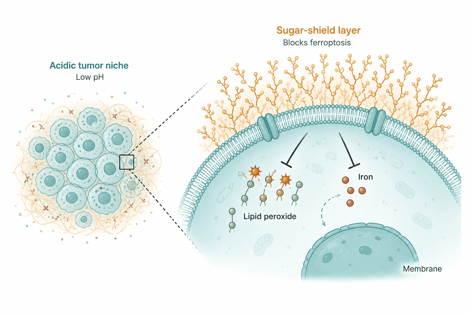
A cluster of tumour cells in an acidic niche, and a magnified stretch of one cell's membrane where a dense glycan shield sits outside and two inhibition bars hold back lipid peroxidation and free iron inside.
Prompt
Create a scientific figure about: Acidic niche sugar shield that blocks ferroptosis. Visual style: Clean Vector. Requirements: publication-ready scientific quality, clear labels and directional arrows where appropriate, avoid cartoon or stock-photo look, composition optimized for research papers and academic slides.
What this template is
The figure argues one chain of cause, from outside the cell to inside it. An acidic tumour niche, shown as a packed cluster of cells in fibrous matrix, is linked through a zoom box to a single membrane. Branched sugar chains crowd its outer face, and from the membrane two blunt inhibition lines reach the two drivers of ferroptosis: oxidised membrane lipids and the pool of free iron. Read from left to right, it moves from tissue to molecule. A reader should come away knowing where the shield sits, which molecules it restrains, and that the cell death it prevents is iron-dependent lipid peroxidation.
Elements to label
Acidic tumour niche
The left-hand cell cluster in fibrous matrix, titled as the tumour niche with low pH as its subtitle.
Zoom box
A small square on the cluster's edge with dashed lines to the right, showing whose membrane is enlarged.
Sugar-shield layer
Dense orange branched glycans above the bilayer; name the actual glycan class if you have measured it.
Phospholipid bilayer
The curved membrane with two transmembrane proteins, which need real names if they stand for transporters.
Lipid peroxides
Phospholipids carrying orange starburst radicals; label them as oxidised lipids, not as generic damage.
Free iron
A few red spheres labelled iron, ideally marked as the ferrous pool that feeds peroxidation.
Inhibition bars
Two T-ended lines from beneath the membrane, one onto the peroxides and one onto iron, drawn in a single style.
Panel heading
The heading above the glycans states the conclusion, that the layer blocks ferroptosis, in slightly larger type than other labels.
Where it is used
- Paper mechanism figure
- Graphical abstract
- Defense slides
- Grant proposal
What reviewers look for
How the shield works
The figure shows two inhibition bars but no mechanism: fewer iron imports, a barrier to the peroxidation chain, or support for a GPX4-type antioxidant axis. A reviewer will ask, so the caption has to say which.
The nucleus is labelled membrane
The label reading Membrane at the lower right points at the nucleus. This is a generation error; relabel it as the nucleus or move the label onto the bilayer before anyone else sees it.
From acidity to glycans
Low pH appears only as text, with no protons, lactate or upstream step showing how acidity builds the glycan layer. If that link is your finding, it needs its own evidence and a visible arrow.
The dashed arrow near iron
A curved dashed arrow runs between the iron and the nucleus, and readers may take it as iron entering the nucleus. Delete it, or state whether it means transport or a signal.
How to use it in Yila
- 1Pick a figure
- 2Describe your subject and the steps to stress
- 3Refine it in chat, then download the PNG
Tips
Do
- Ask Yila to add a ferroptotic endpoint, such as a ruptured cell, so the inhibition bars clearly protect against something.
- If you measured sialic acid or mucins, name the glycans by composition rather than calling them a sugar shield.
- Merge the niche title and its subtitle into one line to give the magnified membrane more room for labels.
Avoid
- Do not keep the Membrane label that points at the nucleus; reviewers read it as a conceptual mistake.
- Do not leave the two membrane proteins as unnamed cylinders, or readers will assume they are the key transporters you claim.
Questions
Can I put this straight into a paper?
Treat it as a first draft of the mechanism panel. It is an AI-generated raster image, and at minimum the nucleus label must be corrected and the shield's mode of action added. After that, keep refining it in Yila until type sizes, line weights and colours meet the submission guidelines of your target journal.
Can elements in the figure be changed?
Not element by element, but you can refine it by chatting in Yila: swap the generic glycans for the ones you measured, name the two membrane proteins, add a GPX4 box below the bilayer, or drop the cell cluster on the left entirely, then generate a fresh version from that description.
What format is delivered?
An AI-generated PNG image with a 4:3 landscape layout. It is not a vector graphic, so the small labels inside the membrane soften when the figure is shrunk; check it at the size it will actually appear in the paper or on a slide.
Is it free?
Viewing this page and the sample figure costs nothing. Generating or revising a version in Yila consumes credits, and a new account receives 2,500 credits, which is enough to fix the labelling problem and work out the mechanism notes over several rounds before you decide on further variants.
I study ferroptosis inducers. Can it show a drug breaking the shield?
Yes. Keep the acidic cluster and the zoomed membrane, ask for the drug and its target to be drawn in, and add a second state where the shield thins and peroxides accumulate. Side by side, the two halves then compare protection with its loss, which is usually the comparison your data actually test.
Can I keep only the magnified membrane?
Ask Yila to remove the cell cluster and the zoom lines and keep only the glycan layer, the bilayer and the inhibition bars. The tighter version leaves room for larger labels and fits a corner of a graphical abstract or a single slide better than the full two-part layout.