Figure template · Mechanism & Pathway · 4:3
Notch Signaling Pathway Mechanism Figure
A landscape mechanism figure in which a signal-sending cell meets a signal-receiving cell, tracing ligand binding, the two proteolytic cuts, NICD entry into the nucleus and target-gene activation, for developmental, stem cell and cancer biology papers.
Prompt
Create a scientific figure about: Notch signaling with Delta/Jagged ligands, proteolytic cleavage, NICD translocation, and target-gene activation. Visual style: Clean Vector. Requirements: publication-ready scientific quality, clear labels and directional arrows where appropriate, avoid cartoon or stock-photo look, composition optimized for research papers and academic slides.
What this template is
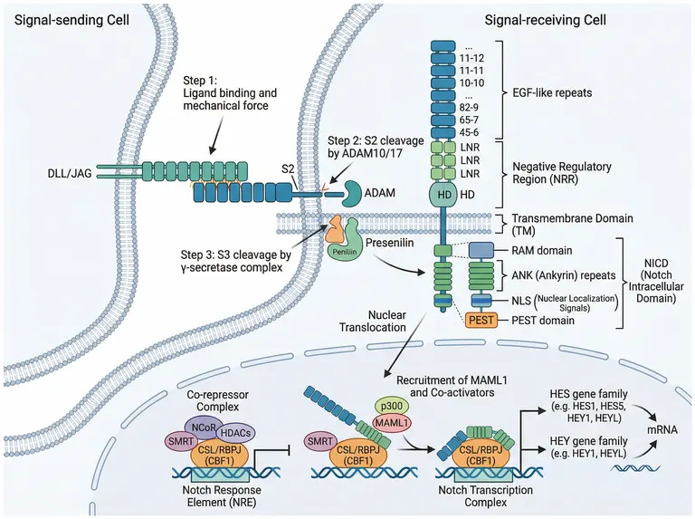
A DLL/JAG ligand on the sending cell grips the EGF-like repeats of a Notch receptor on its neighbour. The pull exposes the negative regulatory region, an ADAM metalloprotease removes the ectodomain at S2, and gamma-secretase then cuts inside the membrane at S3, freeing the Notch intracellular domain with its RAM, ankyrin, NLS and PEST regions. In the nucleus NICD displaces the co-repressor complex from CSL/RBPJ, recruits MAML1 and p300 and switches on HES and HEY genes. A reader should come away knowing which cut comes first and where each one happens.
Elements to label
DLL/JAG ligand
Anchor it in the sending cell's membrane, name the ligand family, and show binding plus the pulling force with arrows.
EGF-like repeats
The stack of extracellular modules; the sample numbers them with nonsense, so label the domain and drop the numbering.
Negative regulatory region
Bracket the LNR modules and HD together as NRR, noting that at rest they shield the S2 site.
S2 cleavage
Mark the juxtamembrane cut by ADAM10 or ADAM17 outside the membrane, numbered separately from S3.
Gamma-secretase and S3
Place the presenilin complex in the receiving cell's membrane; the sample misspells presenilin and drifts it toward the sending cell.
NICD domains
Bracket RAM, ankyrin repeats, NLS and PEST in that order, with the full name of the domain beside the bracket.
CSL switch
Draw the same DNA site twice: first with the co-repressor complex, then with NICD, MAML1 and p300 assembled.
HES and HEY targets
List the two families separately; the sample also puts HEY1 and HEYL under the HES heading.
Where it is used
- Paper mechanism figure
- Course handout
- Grant proposal
- Defense slides
What reviewers look for
The S1 cleavage is missing
During maturation the receptor is cut at S1 by furin in the Golgi, producing the heterodimer the figure implies with its HD label. A paper on receptor processing or trafficking should add that step.
Order and place of the cuts
S2 must precede S3, and S3 happens within the receiving cell's membrane. In the sample the gamma-secretase sits near the junction between the cells, which invites a reader to place it in the sending cell.
What the active complex contains
Once transcription is switched on, co-repressors such as SMRT should have been exchanged out, yet the sample keeps SMRT beside the activating complex. Remove it, or say in the legend that the panel shows a transition.
Ligand endocytosis supplies the force
Endocytosis of the bound ligand is what pulls the regulatory region open. If your paper discusses mechanical activation, add an endocytic vesicle and ubiquitination mark in the sending cell rather than a bare force arrow.
How to use it in Yila
- 1Pick a figure
- 2Describe your subject and the steps to stress
- 3Refine it in chat, then download the PNG
Tips
Do
- Name the receptor paralogue and ligand you study, for example Notch1 with DLL4 or JAG1, so the labels stop being generic.
- If you test gamma-secretase inhibitors, ask Yila to place an inhibition symbol at the S3 site with the drug class written beside it.
- For teaching, request a version with the domain brackets removed so the two cleavages and nuclear entry carry the whole story.
Avoid
- Do not keep the garbled repeat numbering on the ectodomain; ask for domain names only when you regenerate.
- Do not let the three nuclear complexes run together on one strand of DNA without a divider, or repressed and active states blur.
Questions
Can this Notch figure go straight into my manuscript?
Treat it as a first draft of the mechanism panel. It is an AI-generated image whose repeat numbering is garbled, whose presenilin label is misspelled and whose HES list includes HEY genes, so check each label against the literature you cite and keep refining it in chat before submission.
Can I change the ligand to JAG1 and emphasise nuclear entry?
Not element by element, but you can keep refining it by chatting in Yila: ask for JAG1 throughout, remove the domain brackets, enlarge the transcription complexes in the nucleus so they read at slide size, and generate a new version from there.
What format will I download?
An AI-generated PNG image in a 4:3 landscape frame, which fits a figure panel or a slide. It is not a vector graphic, and the small NICD domain labels blur when enlarged, so check them at your final print size before a poster.
Do I have to pay to use it?
Reading this page and viewing the sample cost nothing. Generating and revising in Yila consumes credits, and a new account receives 2,500 credits, which covers several rounds of swapping in your own ligand, receptor and target genes until the panel matches your system.
Can it show cis-inhibition or non-canonical Notch signaling?
Describe what you need in the conversation, such as ligand and receptor in the same cell blocking each other, or a branch that does not run through CSL, and ask for the extra cell and arrows to be added to the existing layout before regenerating.
Is it suitable for teaching lateral inhibition?
Yes. Two touching cells above a nucleus are the natural frame for lateral inhibition and fate decisions in development. For a lecture, ask for a simplified version that keeps the two cleavages and nuclear entry and drops the structural detail.