Figure template · Cross-section · 4:3
Human Skin Cross-Section Anatomy Figure
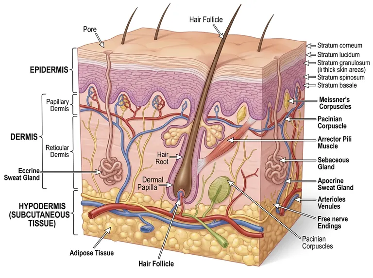
A block of skin cut like a specimen: the epidermal strata are labelled one by one, the dermis is bracketed into papillary and reticular layers holding follicle, glands, arrector muscle and receptors, and fat with paired vessels fills the base.
Prompt
Create a scientific figure about: Human skin cross-section with epidermis, dermis, and appendages. Visual style: Clean Vector. Requirements: publication-ready scientific quality, clear labels and directional arrows where appropriate, avoid cartoon or stock-photo look, composition optimized for research papers and academic slides.
What this template is
The figure lifts out a block of skin so all three levels are visible at once. On top sits the epidermis, labelled downward from the horny surface to the basal layer whose wavy border interlocks with the dermis. The dermis is bracketed into a papillary layer against the epidermis and a deeper reticular layer carrying the hair follicle, its sebaceous gland and arrector muscle, sweat glands and the tactile receptors. Beneath it the fatty hypodermis holds paired arteries and veins that branch upward into the dermis.
Elements to label
Epidermal strata leaders
Lead each stratum out in order from the surface, with thickness and colour showing how keratinisation changes.
Dermal layer bracket
Bracket papillary and reticular dermis on one side, aligning the boundary with the wavy epidermal base.
Follicle and root
The shaft leaves the surface and the root ends in a dermal papilla, the whole follicle slanting through the dermis. Keep the angle consistent.
Sebaceous gland and arrector muscle
Open the gland into the upper follicle and stretch the muscle between follicle and epidermis at an angle.
Two kinds of sweat gland
Coil the eccrine gland deep in the dermis with a duct to a surface pore, and label the apocrine gland apart.
Tactile receptors
Small corpuscles in the dermal papillae, lamellated corpuscles deep down, and free endings reaching the epidermis. Name each receptor once.
Fat and vessels
Draw the base as fat lobules, with arteries and veins running in pairs and branching upward toward the dermis.
Pore and shaft on the surface
Label the sweat pore and the emerging shaft once each, so ducts can be matched to their gland. Keep both on the top face.
Where it is used
- Paper mechanism figure
- Course handout
- Defense slides
- Academic poster
What reviewers look for
Clear layer belongs to hairless skin
The sample draws hair follicles and still lists the stratum lucidum, which appears only in thick palmoplantar skin. Either drop that stratum or move the parenthetical onto it and redraw the block as hairless.
Depth of the lamellated corpuscle
Lamellated corpuscles sit in the deep dermis and hypodermis, yet one leader in the sample lands near the epidermis. Any argument about deep receptive fields fails if that depth is wrong. Place its leader in the deep layers.
Where the apocrine duct opens
Apocrine ducts usually empty into the upper follicle rather than onto the surface, while eccrine ducts open on their own. Draw the two ducts ending in different places. Label both ducts separately.
Duplicated labels and body site
The sample labels the follicle and the lamellated corpuscle twice. Epidermal thickness also varies widely by site, so name the body region in the caption if your text discusses one.
How to use it in Yila
- 1Pick a figure
- 2Describe your subject and the steps to stress
- 3Refine it in chat, then download the PNG
Tips
Do
- If one appendage is your subject, ask for it enlarged as an inset while the other layers stay as outlines.
- Name the cell types you care about in the prompt, such as basal melanocytes or dermal fibroblasts.
- Say which body region the block comes from, since thickness and appendage density change from site to site.
Avoid
- Do not combine thick hairless skin and hairy skin features in one block; their stratification genuinely differs.
- Do not crowd every leader onto one side; split the labels left and right rather than shrinking the type.
- Do not scale the fat layer down to a thin strip; its depth is part of how the three levels read.
Questions
Can this go straight into a paper?
Use it as a draft anatomical figure. Check the stratum names, receptor depths and duct destinations against your own text, then keep refining it in chat until resolution and type size meet the journal's requirements. Name the body site in the caption.
Can elements in the figure be changed?
Not element by element, but you can keep refining it by chatting in Yila: switch to hairless thick skin, add immune cells, drop the receptors and keep only appendages, or pull the vascular network into its own layer. Raise one change per message so versions stay comparable.
What format is delivered?
An AI-generated PNG as a landscape cut block, which suits handouts and posters. It is not a vector graphic, so check that the fine stratum labels remain readable before placing it in a two-column paper. Leaders can also be split to both sides.
Is it free?
Browsing this page and viewing the sample cost nothing. Generating your own version in Yila consumes credits, and a new account receives 2,500 credits, enough to retune the block to your body site and relabel it over several rounds. Group your relabelling requests to spend fewer rounds.
Can disease or wound healing be added?
Yes. Ask for inflammatory infiltrate in the reticular layer, new vessels, or epithelial migration at a wound edge, and those changes are drawn into the matching layers while the stratification is kept. A healthy block can sit beside it for comparison.
Can I keep only the epidermis?
Ask for the epidermal strata alone, enlarged to fill the frame, which suits a lecture on keratinisation; the dermis and fat can shrink into a single band along the bottom. Ask for larger stratum captions there.