Figure template · Lab Apparatus · 4:3
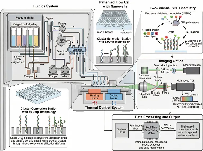
High-Throughput DNA Sequencer Schematic
A whole-instrument schematic with the reagent fluidics on the left, the patterned flow cell and thermal stage in the middle, and two-channel synthesis chemistry, imaging optics and real-time base calling along the right.
Prompt
Create a scientific figure about: Next-generation DNA sequencer with fluidics, patterned flow cell, and dual-channel imaging. Visual style: Clean Vector. Requirements: publication-ready scientific quality, clear labels and directional arrows where appropriate, avoid cartoon or stock-photo look, composition optimized for research papers and academic slides.
What this template is
The figure opens a sequencing-by-synthesis instrument module by module. Chilled reagent cartridges feed pumps, rotary and selection valves and a manifold; single library molecules settle into the nanowells of a patterned flow cell and amplify in place into monoclonal clusters. Each cycle incorporates a nucleotide carrying a dye and a reversible terminator, images the surface, then cleaves both groups before the next round. The imaging optics carry the signal to the camera while heating and cooling blocks drive the temperature, and an on-board processor turns raw images into base calls and output files.
Elements to label
Reagent bay and fluidics
Chain the chiller, cartridge bay, sipper, pumps, valves and manifold, naming which lines carry buffers and enzyme mix along the way.
Patterned flow cell
Section the nanowell array on its glass substrate and keep the spacing between wells visibly even.
Clustering inset
One library molecule entering a well and amplifying in place, with the point that a single clone occupies it.
Synthesis cycle
Close the loop of incorporation, imaging and cleavage, and mark both dye and reversible terminator on the nucleotide.
Imaging optics
Excitation passes through shaping optics and a dichroic into the objective, returning via emission filters to the camera.
Thermal stage
Colour the heating and cooling blocks under the flow cell apart and show heat direction with arrows.
Data path
Line up the on-board processor, real-time base calling and the output module, naming the files handed on at each step. Keep that row horizontal.
Waste return
End the fluidics at waste, drawing the return line thinner than the reagent supply so the two do not merge visually.
Where it is used
- Paper mechanism figure
- Defense slides
- Course handout
- Lab website
What reviewers look for
How two channels encode four bases
Two channels distinguish four bases only because one base lights both and one lights neither. The sample prints four letters and two dyes, so add a small table of the signal combinations beside the cycle.
Clusters must be monoclonal
Two different molecules in one nanowell give a mixed read. The clustering inset should show a single clone holding the well and name the exclusion mechanism that keeps it that way. Say it in the caption as well.
Imaging synchronised with motion
The camera caption in the sample is garbled. Charge transfer has to track flow cell movement or the scan smears, and that sentence is what explains the instrument's speed.
Incomplete cleavage causes phasing
Leftover dye or terminator leaves strands a cycle behind and quality drops along the read. If your text discusses phasing, keep a branch in the cycle for strands that failed to cleave. Label that branch plainly.
How to use it in Yila
- 1Pick a figure
- 2Describe your subject and the steps to stress
- 3Refine it in chat, then download the PNG
Tips
Do
- Keep only the modules your talk needs; a library-methods figure can hold the flow cell and cycle and drop the plumbing.
- State the read structure, so the cycle panel can show single or paired end reads and any index reads.
- Pull leader lines horizontally from each module edge; crossing diagonals make a whole-instrument view unreadable.
Avoid
- Do not print a model name or a vendor on the figure; instrument identity belongs in the methods text.
- Do not let one module appear twice with its own caption, which reads as two separate pieces of hardware.
- Do not stretch the fluidics across half the frame unless plumbing is your subject; the chemistry panel carries the science.
Questions
Can this go straight into a paper?
Use it as a draft instrument figure. Check the module names, repair the garbled captions, make sure no model name survived, and keep refining it in chat until the resolution and type size suit the journal. Line weights matter at this density.
Can elements in the figure be changed?
Not element by element, but you can keep refining it by chatting in Yila: remove the data path, switch to a four-channel chemistry, keep only the flow cell section, or add library preparation upstream, then generate again. Ask for one change at a time so each version stays comparable.
What format is delivered?
An AI-generated PNG in a landscape frame with leader lines between modules. It is not a vector graphic, and the whole-instrument view is dense, so check the smallest captions before reducing it to column width. A two-row arrangement helps when the figure must sit in one column.
Is it free?
Browsing this page and viewing the sample cost nothing. Generating your own version in Yila consumes credits, and a new account receives 2,500 credits, enough to cut the schematic down to the modules you need over several rounds. Group the edits you want before generating a new version. Fewer rounds means fewer credits spent.
I work with long-read sequencing. Can I reuse it?
The layout carries over, but the chemistry block must be replaced: swap clustering and reversible termination for single-molecule real-time signal or pore current. Fluidics, thermal control and the data path keep their places. Say which readout you use when you ask.
Can I keep only the synthesis cycle?
Yes. Ask for the incorporation, imaging and cleavage loop alone, enlarged to fill the frame for a chemistry lecture, with the remaining modules either shrunk into one strip or dropped entirely. Larger nucleotide labels are worth asking for there.