Abbildungsvorlage · Mechanismus und Signalweg · 4:3
Mitochondrial Transplantation Eye Cutaway Figure
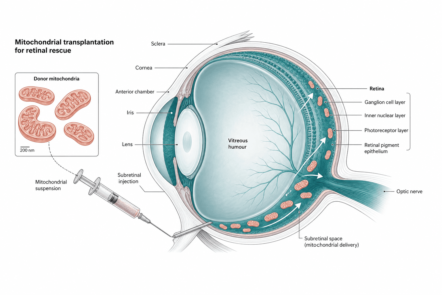
A sagittal cutaway of the eye with a syringe entering through the sclera, white arrows tracing where the delivered mitochondria spread through the retina, and an inset enlarging the donor organelles beside a scale bar.
Prompt
Create a scientific figure about: Organelle-delivery cutaway for transplantation. Visual style: Clean Vector. Requirements: publication-ready scientific quality, clear labels and directional arrows where appropriate, avoid cartoon or stock-photo look, composition optimized for research papers and academic slides.
Was diese Vorlage ist
The figure answers how the cargo travels. Donor mitochondria are prepared as a suspension, drawn into a syringe, pushed through the sclera into the space beneath the retina, and then distributed through the retinal layers. The cutaway supplies the anatomy, with leader lines naming the cornea, iris, lens, vitreous and optic nerve, while a bracket on the right breaks the retina into ganglion cell layer, inner nuclear layer, photoreceptors and pigment epithelium. A reader should be able to restate where the needle enters, which space the suspension fills, and which cells the organelles are meant to reach.
Zu beschriftende Elemente
Sagittal eye cutaway
The main body keeps anterior and posterior segments, cut in one direction, with the wall coloured apart from the retina.
Syringe and needle track
The needle crosses the sclera at an angle and stops under the retina without covering the lens or the posterior pole.
Donor mitochondria inset
A boxed enlargement of several cristae-bearing organelles with a scale bar, tied to the eye by a dashed line.
Retinal layer bracket
A bracket listing the four layers from inner to outer, each with its own leader line rather than one shared anchor point.
Delivery arrows
White curved arrows inside the retina point towards the photoreceptors; keep one arrow style for one meaning.
Anatomical labels
One leader each for cornea, iris, lens, vitreous, sclera and optic nerve, with the text set outside the globe.
Corner title
Two short lines of title above the inset, naming the organelle being delivered and the tissue it is meant to rescue.
Wofür sie verwendet wird
- Paper mechanism figure
- Graphical abstract
- Defense slides
- Grant proposal
Worauf Gutachter achten
Needle tip must match the label
The label says subretinal injection while the tip sits near the ciliary body and vitreous border. If your route is really intravitreal or suprachoroidal, move the tip or change the wording.
The spread is schematic
Organelles appear evenly spread across the whole retina, whereas uptake in practice concentrates near the injection site. State in the caption that the distribution is illustrative rather than quantitative.
Layer names and order
From the vitreous outward the bracket should read ganglion cell layer, inner nuclear layer, photoreceptors and pigment epithelium. A reversed order or a missing outer nuclear layer is the first thing a retina reviewer notices.
Uptake is not shown
Nothing in the figure explains how recipient cells internalise the organelles, or whether membrane potential survives the transfer. If that is part of your claim, it needs its own inset and its own evidence.
So verwenden Sie die Vorlage in Yila
- 1Abbildung auswählen
- 2Forschungsgegenstand und zu betonende Schritte beschreiben
- 3Im Chat weiter verfeinern und dann als PNG herunterladen
Tipps
Empfehlenswert
- Draw the organelles inside the retina exactly as they appear in the inset so readers recognise them as the same cargo.
- Put a scale bar on both the inset and on one retinal detail, so the size difference between them is immediate.
- Name the target space in the prompt and insist that the needle tip stops precisely there, not nearby.
Besser vermeiden
- Do not run leader lines across the inside of the globe to reach outer structures; crossing leaders always cost this kind of cutaway marks.
- Do not colour the retina and choroid alike, or no reader can tell which layer the arrows actually end in.
Häufige Fragen
Can this go straight into a paper?
Start from it as an anatomical sketch. It is an AI-generated raster image, so check that the needle tip really sits in the space the label names, rewrite the spread of organelles to match what you observed, and then keep refining line weights and type sizes in Yila until they suit your target journal.
Can elements in the figure be changed?
Not element by element, but you can keep refining it by chatting in Yila: switch the route to an intravitreal injection, add an inset showing uptake by a recipient cell, or drop the anterior segment and keep only the posterior pole, then generate a new version from that description.
What format is delivered?
An AI-generated PNG image in a 4:3 landscape layout. It is not a vector graphic, so the four stacked retinal labels blur together once the figure is reduced; check it at your actual column width before using it as a paper figure.
Is it free?
Viewing this page and the sample costs nothing. Generating or revising a version in Yila consumes credits, and a new account receives 2,500 credits, enough to iterate on the injection route, the layer labels and the inset position until they read correctly.
I deliver into brain tissue, not the eye. Can it be adapted?
Yes. The skeleton is an organ cutaway, a delivery route and a layered target, so replace the globe with a brain section and the retinal bracket with cortical layers. The inset and the scale bar work in exactly the same way in both versions.
Can I keep only the organelle inset?
Ask for the donor mitochondria and the scale bar alone, without the globe and its leader lines. On its own that panel suits a corner of a graphical abstract or a methods figure, where its job is to document the shape and the size of the material you are delivering.